Friday, November 2, 2018

Oral Cancer and HPV

In our efforts to provide the highest standard of care our team routinely attends continuing education courses.  Our hygienists recently attended an update in Oral Cancer. The presentation was given by the surgeons from Washington University School of Medicine, Department of Head and Neck Surgical Oncology. Dr. Jose P. Zevallos, the Division Chief  started the morning with  an update on oropharyngeal cancer.

Dr. Zevallos recounted the rise in orophoharyngeal cancer began to be noticed in the 1980's and 1990's. It wasn't until the early 2000's it was discovered that this rise in cancer was being caused by  HPV (Human papillomavirus). The scary thing about this cancer is that it's demographic is across all age ranges, in smokers and non-smokers, with an increase noted in all races. Another troubling thing about HPV related cancers is that it has a 30 year legacy period between infection and onset of cancer. The infection generally occurs in the patients 20's and then lays dormant until the 50's to 60's. People with autoimmune diseases or people taking immunosuppressants are at a higher risk for developing HPV related cancers.

Another issue associated with HPV oropharyngeal cancers is our lack of ability to reliably screen for them. There are some saliva tests for oral HPV infection, however there is a high rate of false positives with these tests. This is because 2-8% of the population will have the infection but only a tiny percent of those get cancer. Once a tumor is present there will be very high levels of HPV in the saliva. The current saliva tests do not screen for the levels of HPV present in the saliva. Secondly, these tumors form deep in the base of the tonsillar crypt and the base of the tongue, making them very hard to see in a visual oral exam.

The Gardisil vaccine now cover 9 of the high risk forms of HPV. This vaccine is our best chance at preventing cancers. However, much of the population is still at risk for developing an HPV related cancer of the head, neck and mouth. This is why it is critical to have regular dental checkups and ensure your provider is competently evaluating your oral and oropharyngeal cavities for any early signs of cancer.


Possible signs and symptoms of oral cavity and oropharyngeal cancers include:
  • A sore in the mouth that doesn't heal (the most common symptom)
  • Pain in the mouth that doesn’t go away (also very common)
  • A lump or thickening in the cheek
  • A white or red patch on the gums, tongue, tonsil, or lining of the mouth
  • A sore throat or a feeling that something is caught in the throat that doesn’t go away
  • Trouble chewing or swallowing
  • Trouble moving the jaw or tongue
  • Numbness of the tongue or other area of the mouth
  • Swelling of the jaw that causes dentures to fit poorly or become uncomfortable
  • Loosening of the teeth or pain around the teeth or jaw
  • Voice changes
  • A lump or mass in the neck
  • Weight loss
  • Constant bad breath
Many of these signs and symptoms can also be caused by things other than cancer, or even by other cancers. Still, it's very important to see a doctor or dentist if any of these conditions lasts more than 2 weeks so that the cause can be found and treated, if needed.

Friday, October 12, 2018

Oral Bacteria Use Your Bloodstream As Highway To Your Body

PERIODONTAL PATHOGENS AREN'T JUST IN YOUR MOUTH


The concept of sterile blood no longer exists. Bacteremia occurs when pathogens enter the bloodstream. Many microbes simply enter the bloodstream transiently. They are shed from the primary biofilm found in the mouth by chewing or routine dental hygiene at home.
Higher concentrations of pathogens enter the blood by invasive dental procedures, including mechanical debridement, extractions, and endodontic procedures. It requires only 60 seconds for a pathogen to travel anywhere in the body as a result of an invasive dental procedure. 
Bacteremia results in “seeding” bacteria at potentially new systemic locations. These new sites are typically under stress and have become susceptible to invasion. 
Translocation is a pathogen’s arrival at a new systemic site from its point of origin. New biofilms or microbiomes of pathogens emerge at sites where these species are not normally found. The impact of these pathogens is the same wherever they colonize: cellular destruction.
Co-Occurrence
In the past several years, much evidence has shown a prevalence of periodontal pathogens in arterial biofilms and microbiomes. Recently, the term “co-occurrence” has become relevant as it describes the existence of two similar biofilms in the same patient at the same time.
study published in February 2017 found that a large group of patients who had both the symptoms of periodontitis and of heart disease had a similar profile of periodontal high-risk pathogens at the two independent locations. For three pathogens, the co-occurrence was at the 95% confidence level. Eight other pathogens were simply determined to have a very high incidence of correlation. 
In a dental practice, a simple salivary test can be performed to determine the specific bacteria that are present in the mouth, understanding that the very same bacteria that are found in high concentrations in the mouth also are found on the lining of the patient’s arterial walls.
The goal of periodontal therapy should be to eliminate the bacterial etiology. Successful periodontal therapy must include antimicrobials and or antibiotics specific to the pathogens present in a given individual’s oral bacterial flora or biofilm. 
If periodontal therapy is only delivered with a local focus, what will become of the already translocated and in-place biofilms and pathogens in hosts who are medically or genetically compromised and incapable of mounting an effective immune response? Does a systemic disease warrant a systemic response? These questions need to be considered when periodontal therapy is warranted.
Looking at periodontal disease as a bacterial infection that travels throughout the body is much different than looking at periodontal disease as a localized bacterial infection of the oral cavity only. Systemic mitigation of pathogens requires a total body wellness approach. Periodontal disease treatment must be considered a part of the preventive armamentarium for chronic disease management in the human body, which is a complex ecosystem. 
This article excerpted from:
Dentistry Today
08 Oct 2018  Traci Warner, RDH  Today's Dental News

Friday, October 5, 2018

New Research: Smoking Can Damage Your Tooth's Nerve


Smoker's have been cautioned about lung disease, gum disease and other deleterious affects of smoking, but a new study has shown that smoking can affect the pulp (nerve and blood supply inside your tooth) of your tooth.
Researchers at the Case Western Reserve University School of Dental Medicine found that smoking weakens the ability for pulp in teeth to fight illness and disease.
In other words, smokers have fewer defense mechanisms on the inside of their teeth.
"That might explain why smokers have poorer endodontic(root canal treatment) outcomes and delayed healing than non-smokers," said Anita Aminoshariae, associate professor of endodontics and director of predoctoral endodontics. "Imagine TNF-? and hBD-2 are among the soldiers in a last line of defense fortifying a castle. Smoking kills these soldiers before they even have a chance at mounting a solid defense."
The results of the study were published in the Journal of Endodontics.
Aminoshariae said that, previously, there was little research into the endodontic effects of smoking -- the inside of teeth. Smokers had worse outcomes than nonsmokers, with greater chances of developing gum disease and nearly two times more likely to require a root canal.
This new preliminary research set out to explain the possible contributing factors.
Thirty-two smokers and 37 nonsmokers with endodontic pulpitis -- more commonly known as dental-tissue inflammation -- were included in the study.
"We began with a look at the dental pulp of smokers compared with nonsmokers," she said. "We hypothesized that the natural defenses would be reduced in smokers; we didn't expect them to have them completely depleted."
One interesting find, Aminoshariae noted, was that for two patients who quit smoking, those defenses returned.
Joining Aminoshariae in the study were former students Caroline Ghattas Ayoub and Mohammed Bakkar; faculty members Tracey Bonfield, Catherine Demko, Thomas A. Montagnese and Andre K. Mickel; and research Santosh Ghosh -- all from the School of Dental Medicine.

Case Western Reserve University. (2018, September 26). Dental research shows that smoking weakens immune systems. ScienceDaily. Retrieved October 5, 2018 from www.sciencedaily.com/releases/2018/09/180926110940.htm

Friday, August 17, 2018

Plaque: The invisible enemy of teeth

The CONSTANT BATTLE WITH BACTERIA


We come into the world without teeth. Too often we exit in the same condition.
That’s an odd outcome, given that enamel, the white part of the tooth, is the hardest substance in the body. Human teeth are actually as hard as sharks’ teeth, a group of scientists from Germany discovered. Sharks have advantages over us, though. Rows of sharks’ teeth are replaced throughout life, while loss of a permanent human tooth requires a prosthesis in the form of a bridge, crown, implant or dentures.
If our teeth are so strong, why then are 19 percent of the population 65 years old and older completely edentulous, or toothless?
Sharks again provide part of the answer. They don’t get cavities — and with good reason. Sharks don’t eat sugar, but we do, and apparently, plenty of it. Added sugar, such as table sugar, honey and high fructose corn syrup, are the major culprits here, but all sugars and starches play a role. The World Health Organization recommends no more than 25 grams of added sugar a day, which amounts to 20 pounds a year. Yet, the average American consumes 82 grams a day, or 66 pounds a year.
illustration: Caleb Olson

Plaque — step one to cavities

The teeth are covered by plaque a soft, invisible sticky film teeming with bacteria – some good, some bad. Plaque is always in the mouth even after you brush. That’s because bacteria are hardy creatures and multiply very rapidly. The problem begins when you start to eat. When you eat sugars or any carbohydrate or starch, like rice and bread, the bad bacteria start eating too. They feed off sugars, and that combination of sugar and bacteria is not a good thing. It produces an acid which starts the destructive process of tooth decay called caries or cavities.
The problem is that plaque will not remain soft and sticky. If not brushed away after a period of time, it turns into a hard yellowish substance called tartar, which can be removed only by a dentist or dental hygienist.
Dental caries is very common in this country. Approximately 91 percent of U.S. adults aged 20 to 64 had dental caries in permanent teeth, according to a 2015 data brief by the National Center for Health Statistics. Apparently, this process starts at an early stage. More than 43 percent of youth between the ages of two and 19 had both treated and untreated cavities in primary or permanent teeth.
Minorities are hardest hit. Blacks, Hispanics and Native Americans have the poorest oral health of any racial and ethnic groups in the United States. In addition, less educated adults experience untreated tooth decay nearly three times that of adults with at least some college education

Gum disease

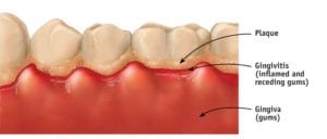
Plaque is not confined to the teeth. It can also initiate an infection of your gum tissue and bone surrounding your teeth. The initial stage of gum disease  is called gingivitis, which is characterized by redness and bleeding gums particularly when brushing or flossing. With good oral care, this stage is reversible. It can progress, however, to periodontitis, which is irreversible and can result in permanent tooth loss.
Image: courtesy of Mayo Clinic
What’s worse is that the inflammation can have an impact on chronic diseases, such as diabetes and heart disease. “The mouth is part of the body,” said Dr. Susana Tejada, a dentist at The Dimock Center. “Bacteria that affect the mouth can travel to the rest of the body. It’s all connected.”
Research has found that plaque builds up in people with diabetes that is poorly controlled. “People with diabetes are three to four times more likely to have gum disease,” she explained. The reverse is also true. Gum inflammation makes it harder to control diabetes.
It’s not just diabetes that’s affected by poor oral health. The heart may suffer as well. Some studies suggest that clogged arteries that contribute to heart disease and stroke may have a connection to gum inflammation, according to the Mayo Clinic. Women with periodontitis have a higher risk of premature birth and low birth weight.

Dry mouth

Saliva — known more familiarly as spit — does not have the best reputation. Your mother taught you not to spit. Several states — including Massachusetts — have laws on the books forbidding residents to spit on a public sidewalk. In the Commonwealth, that antiquated law carries a whopping $20 fine. These laws and your mother’s admonition give the impression that there’s something sinister about spit. Yet, saliva is nourishing to the mouth. “It washes away food particles and moistens the mouth,” explained Tejada. “It prevents tooth decay by neutralizing the acids produced by bacteria.”
Low production of saliva results in a condition known as dry mouth. Patients complain that it feels like “cotton balls in the mouth,” or “everything seems to stick.” Dry mouth is often a side effect of certain medications, such as those to treat depression, high blood pressure, and mental health disorders anxiety and pain. Even over-the-counter drugs, including antihistamines and decongestants can cause the condition. It is more common in older people, but more probably a factor of increased use of medication than passing years. Therapy for head and neck cancers, tobacco, alcohol and marijuana use are other possible causes.

Types of dentists

General dentist
General dentists are your primary dental providers who manage your overall dental health. They provide cleanings and x-rays, diagnose dental conditions and perform several dental procedures, including fillings and extractions. At times, however, a specialist is required.
Endodontistspecializes in root canals
Oral surgeonperforms complex extractions and surgeries on the mouth and jaw
Orthodontistdevelops braces or other devices to straighten teeth
Pedodontist or pediatric dentistspecializes in the prevention, diagnosis and treatment of diseases that affect the gums, such as gingivitis and periodontitis
Prosthodontistresponsible for replacement of missing teeth and the repair of a person’s natural teeth by means of bridges, crowns, implants and dentures

Dry mouth is more than just annoying. A more serious complication is its link to increased plaque and tooth decay. There’s no moisture to wash away the food, and therefore no protection of the teeth from acid formation. Roughly 30 percent of all tooth decay in older adults is caused by dry mouth, according to the Oral Cancer Foundation.
Over-the-counter remedies such as mouth sprays and sugar-free gum, are available to reduce the symptoms. Tejada offers an easier and free solution. “Drink a lot of water” she advised. “Sip it frequently throughout the day.”

Prevention

Yet, tooth decay and gum disease are largely preventable. You need a toothbrush, floss and fluoride toothpaste … and you have to use them. It’s easy on the budget. All together that can set you back roughly $10 every three months. Check-ups with the dentist can help spot a problem before it balloons into a more serious condition. In spite of this relatively low cost at-home treatment, cavities remain one of the most common chronic diseases in the U.S.
More than 70 percent of the residents in Massachusetts receive fluoridated water. Studies have shown that people who drink fluoridated water have a lower incidence of tooth decay. Fluoride replaces minerals in the teeth that are lost due to acid formation by bacteria.
We can drink the water, but it is necessary to do more than just imbibe and brush. Other habits do their fair share of damage. Smoking decreases the production of saliva, which leads to dry mouth, and in turn the risk of gum disease. But it’s the diet that causes the most problems, and two of the biggest offenders are soda and coffee. Roughly half of the U.S population drinks an average of three glasses of sodas every day. In addition, the average coffee drinker sips two to three cups a day, but coffee is very acidic. That means we are constantly coating our teeth with both sugar and acid, an ill-fated duo.
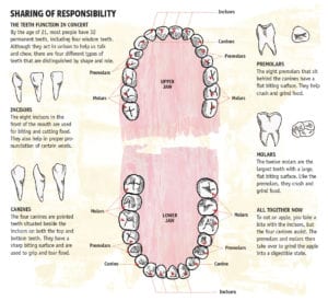
By age 21 we have 32 teeth. At 81 we should still have 32 teeth. Although, due to evolutionary changes, many people never develop their 3rd molars (wisdom teeth) or their jaws are too small to accommodate them and they are extracted, making the total tooth count 28 teeth. Tooth loss is not inevitable with age, warned Tejada. “They do not have to go hand in hand.”
 Article from The Bay State Banner; Be Healthy  by Karen Miller 7-20-18

Friday, August 10, 2018

Oral Hygiene and Type 2 Diabetes

Better Dental Hygiene Linked to Improved Type 2 Diabetes Glycemic Levels


A study published in The Journal of Clinical Periodontology revealed a potential link between Type 2 diabetes and dental hygiene. The results of the study will have a significant impact in the medical community, especially because Type 2 diabetes accounts for approximately 90% of all diabetes cases and impacts over 30 million Americans according to the Center for Disease Control.
A randomized clinical study conducted by researchers at the Faculty of Medicine and Health Sciences of the University of Buffalo demonstrated that patients who follow proper dental hygiene routines might experience an improvement in their Type 2 diabetes. The lead researcher, Miquel Viñas, is a professor of Microbiology at the University of Buffalo. Professors Elisabet Mauri Obradors, Albert Estrugo and Enric Jané from the university’s Department of Odonto-Stomatology also contributed to the study. Additional research participants included Alexandra Merlos from the Department of Pathology and Experimental Therapy and José López López, a lecturer and medical director at the Dental Hospital of the University of Buffalo.

Type 2 Diabetes Causes and Symptoms 

Insulin is a naturally occurring hormone secreted by the pancreas. Type 2 diabetes is a chronic condition which occurs when the body cannot metabolize blood sugar and becomes resistant to insulin. As a result, blood sugar levels can rise higher than normal. This can lead to serious health problems such as kidney and heart disease and other medical complications. People who are obese, and not physically active, may be at risk for developing Type 2 diabetes. Common symptoms include fatigue, frequent urination (polyuria), weight loss, increased thirst (polydipsia) and hunger (polyphagia), blurry vision and thick darkened skin. However, some patients may never experience noticeable symptoms and do not discover they have the disease until their blood sugars are tested, and a physician diagnoses them.
Although Type 2 diabetes is incurable, it can go into remission when patients begin to eat healthy and work out to maintain their blood sugar levels. Some patients may need to take prescribed oral medications or receive insulin injections to help keep their target blood glucose at healthy levels.

The University of Buffalo Study

The researchers decided to evaluate the effect of non‐surgical periodontal treatment on serum HbA1c (hemoglobin A1c or glycated hemoglobin) levels in patients who were diagnosed with Type 2 diabetes. Although previous research had shown a correlation between diabetes and periodontal diseases, the researchers noted there were limited studies about how periodontal disease can affect patients with diabetes.
Ninety patients with Type 2 diabetes participated in the single‐masked, randomized clinical study. Over the duration of six months, patients received oral treatments and underwent a control of glycated hemoglobin. The researchers also evaluated the patients for certain types of oral bacteria that are linked to periodontitis. The treatment group received oral hygiene instructions and non-surgical periodontal therapy/SRP using both ultrasonic and hand instrumentation. Meanwhile, the study’s control group received just oral hygiene instructions and had biofilm and calculus removed with only ultrasonic instrumentation. The researchers measured and analyzed the patient’s plaque index, pocket depth, and gingival index at the beginning, middle, and end of the study.

The Results of the Study

Based on their findings, non‐surgical periodontal treatment resulted in a better glycemic status for patients with type 2 diabetes. “The main conclusion of the study,” José López López shared, “is that the non-surgical treatment of periodontitis improves the glycemic status and levels of glycated hemoglobin, and therefore proves the great importance of oral health in these patients.” There was no noticeable improvement in the control group. In conclusion, the study revealed just how much oral health is closely tied to a patient’s overall health.

Published by Today's RDH August 3, 2018

Friday, July 27, 2018

HAPPY SUMMER

Summer Vacation is Here, Yeah!


We are off for the one week when the entire staff takes a summer vacation.  The staff is off to many different areas of the country with family and friends. 

We will return on Monday August 6th!

Happy Summer To You All

Monday, July 16, 2018

Periodontal Disease Just May Be Causing Rheumatoid Arthritis


BACTERIA THAT CAUSES ADVANCED GUM DISEASE FOUND IN LARGE AMOUNTS IN PATIENTS WITH RHEUMATOID ARTHRITIS


Individuals at risk of rheumatoid arthritis (RA) have increased levels of gum disease and disease-causing bacteria, according to a study presented at the Annual European Congress of Rheumatology (EULAR 2018).
“It has been shown that RA-associated antibodies, such as anti-citrullinated protein antibodies, are present well before any evidence of joint disease. This suggests they original from a site outside of the joints,” said lead author Kulveer Mankia, MD, of the Leeds Institute of Rheumatic and Muscoskeletal Medicine and the Leeds Biomedical Research Centre. 
“Our study is the first to describe clinical periodontal disease and the relative abundance of periodontal bacterial in these at-risk individuals. Our results support the hypothesis that local inflammation at mucosal surfaces, such as the gums in this case, may provide the primary trigger for the systemic autoimmunity seen in RA,” Mankia said.
RA is a chronic inflammatory disease that affects the joints, causing pain and disability. It also can affect internal organs. While it is more common in older people, there also is a high prevalence in young adults, adolescents, and even children, and it affects women more frequently than men. 
Porphyromonas gingivalis
The prevalence of gum disease is increased in patients with RA and could be a key initiator of RA-related autoimmunity. This is because autoimmunity in RA is characterized by an antibody response to citrullinated proteins, and the oral bacterium Porphyromonas gingivalis (Pg) is the only human pathogen known to express an enzyme that can generate citrullinated proteins.
“We welcome these data in presenting concepts that may enhance clinical understanding of the key initiators of rheumatoid arthritis,” said Robert Landewé, MD, PhD, chair of the Scientific Programme Committee with EULAR. “This is an essential step towards the ultimate goal of disease prevention.” 
In the study, dentists diagnosed clinical gum disease in significantly more at-risk individuals than in healthy controls (73% versus 38%). Also, the percentage of sites with clinical attachment level of ≥2 mm, pocket depth of ≥4 mm, bleeding on probing, periodontal disease (PDD), and active periodontal disease were all significantly greater in the at-risk individuals compared to controls. In non-smokers, PDD and active PDD were more prevalent in at-risk individuals compared to controls.
DNA was isolated from the subgingival plaque, next to the gums, of each participant and used to measure the levels of three types of bacteria, PgAggregatibacter actinomycetemcomitans (Aa), and Filifactor Alocis. Results showed that there was increased abundance of both Pg and Aa in at-risk individuals. But in at-risk individuals, only Pg was significantly increased at healthy dental sites and was associated with the overall extent of gum disease.
The study included 48 at-risk individuals (positive test for anti-citrullinated protein antibodies, musculoskeletal symptoms but no clinical synovitis), 26 patients with RA, and 32 healthy controls. The three groups were balanced for age, gender, and smoking. At-risk individuals underwent ultrasound assessment to assess for subclinical synovitis. Only two were found to have ultrasound synovitis. Dentists examined six sites per tooth in each participant, and a clinical consensus was reached in each by three dentists.

02 Jul 2018  Dentistry Today Industry News

Friday, June 29, 2018

The Dangers of Oral Piercings


Today's article is from studies that were recently presented at the European Federation of Periodontology’s EuroPerio9 conference, June 20 to June 23 in Amsterdam.  
Though considered trendy and fashionable in some circles, tongue piercings may affect nearby gums and teeth.  We have seen multiple cases in our practice of  severe damage to the periodontium from these fashion statements. In some cases the damage occurred very quickly, within the first months of the piercing.  
STUDY ONE
“My interest in piercings began when a young female patient presenting unusually severe periodontal destruction was referred to my clinic,” said Clemens Walter, DMD, of the Department of Periodontology, Endodontology, and Cariology at the University of Basel in Switzerland and author of the first study.
“The patient was periodontally healthy with the exception of the lower incisor teeth. We could not identify any known risk factors. The patient had a tongue piercing. Nonsurgical and advanced surgical treatment was performed and was not successful. The patient lost teeth. Since then, I have been curious about this association and started to collect cases,” Walter said.
The 18 subjects in Walter’s study, including 14 females, were drawn from a database of more than 1,400 patients treated at the university. The mean age was 28.3 years, ±7.7 years. Three of the patients wore both a lip and a tongue piercing, for a total of 14 tongue piercings and seven lip piercings.
Clinical parameters and the maximum wearing time of the lip and/or tongue piercing were recorded. Periodontal findings in the teeth close to the piercing were compared to teeth not affected by the piercing.
“Due to ethical considerations, it seems impossible to conduct a prospective study. Therefore, the suitable design to answer our question was a retrospective study/case series,” said Walter.
“We found an association between oral piercings and increased periodontal inflammation, as evident by increased bleeding on probing and increased probing depth and/or attachment loss. The closer teeth were to a tongue piercing, the more affected they were,” said Walter. 
recession and bone loss caused by a lip piercing
“Acknowledging the growing data from all over the world, we now try to create awareness about the consequences of piercings for oral health, and we counsel our patients to remove piercings in order to decrease the risk for dental and periodontal complications,” said Walter.
STUDY TWO
Dr. Bernard Loir of Brussels found repeated gum damage due to the chronic pressure of metal against teeth and gums during tongue movements in speaking, eating, or swallowing in a pair of female patients, age 27 and 32. 
This gum damage also was associated with gum bleeding and infection, causing deep lingual infrabony lesions and periodontitis. The tongue piercings were used for eight to 10 years and finally removed after abscesses, teeth mobility, and migration.
“The lesions were localized on the lower incisor’s lingual side, close to a tongue piercing (the most frequent oral localization of these devices, followed by the lip),” said Loir.
“Periodontal complications occurred in a location hardly visible for the patient, and as these teeth have very short roots, tooth mobility and tooth loss in the aesthetic area may be quickly encountered, an uncomfortable situation for such young and mostly female patients,” said Loir. 
“For both patients, time consuming and costly surgical treatment and antibiotics were needed, and complete regeneration of the lost tissue could not be achieved,” Loir said.
“Young patients with oral piercings should be made aware of the local and global health risks they take by wearing these useless devices, especially if they compound the risk by smoking and a lack of proper dental hygiene. Piercings interfere daily with chewing, swallowing, and speaking, and the rate of complications is very high in the available literature,” said Loir. 
“Early complication is usually infection due to lack of hygiene in the piercing saloon or when done by the patients themselves with nonsterile material. Bleeding and swelling of the tongue is also very common and may represent a danger for a patient with cardiovascular disease, but late complications also occur, such as swallowing the jewelry, teeth chipping, and gum recession with lip piercings,” Loir said. 
“Other complications occur in a more silent mode, such as lingual gum recession and bone loss, causing tooth mobility, migration, and loss. Other life-threatening diseases such as viral contamination (hepatitis, papilloma virus), endocarditis, and cerebral abscesses have also been registered,” said Loir.
“While awareness with respect to oral complications associated with piercings seems to be slowly increasing, there is yet much to be done,” said Walter. “We are continuing with our research and hope to recruit more patients in order to increase our database.”

CONCLUSION
My advice to all people considering a perioral or tongue piercing is very simple, just don't do it. And for all those who have already taken the leap and had the piercing done, your best bet it to promptly remove it. If you've noticed any changes in the condition of your gum near your piecing make an appointment with a periodontist to get the damaged treated, it might not be too late to save the affected teeth.


Friday, June 22, 2018

Possible Gene found which controls periodontal disease

Study pinpoints genetic connections to perio and coronary artery disease in addition to lifestyle factors


Results of a meta-analysis presented at the European Federation of Periodontology-EuroPerio9 meeting reported that periodontal disease and coronary artery disease (CAD) share a common genetic basis. 
“Knowledge of the shared genetic basis helps to understand the molecular mechanisms that underlie and predispose to the disease,” explained lead author, Dr. Arne S Schäfer, a professor at the department of periodontology and synoptic dentistry based at Charité University Medicine in Berlin, Germany. “This knowledge will guide strategies for therapy but also allow the identification of risk groups for preventive care, before the disease manifests itself.”
Strong evidence of association between CAD and periodontal disease (PD) has already been established. Both are among the most common diseases: CAD affects 110 million people worldwide and is the first cause of death, while PD affects 538 million people.(2) Both diseases are frequently diagnosed together and have common risk factors, such as smoking and diabetes. Both are characterised by a chronic inflammatory process but, independent of those shared risk factors, previous studies(3,4) had suggested a few shared genetic variants.
Dr. Schäfer said, “Our aim in undertaking this study was to further explore the joint genetic basis of CAD and PD. The identification of the shared genetic susceptibility factors will pinpoint relevant molecular pathways for the disease. This knowledge will yield very specific therapeutical targets for precision medicine. We believed that, given the localized nature of periodontitis which is confined to the oral cavity, there would be a small variety of different pathways that had the potential to contribute to both diseases.”
He added that the 10-year study examined every common variant in the entire DNA sequence. “These are alternative building blocks called alleles, which are in the millions. We counted if a variant was more common in both CAD and periodontitis cases, compared to healthy controls,” he said. To rule out chance findings, which can be caused by random differences of natural variation, we counted all these variants in all patients of CAD and periodontitis that were available to us. This high number of analyzed individuals and a replication of the results in an independent sample of cases and controls, allows to generalize our findings.”
Researchers identified a variant in the promoter region of the gene VAMP8 (a promoter regulates the activity of a gene in response to other stimuli) to be significantly more frequent in CAD and periodontitis cases than in healthy controls, indicating the involvement of this gene in the etiology of both diseases.
“VAMP8 is of special interest, because it is involved in the import and export of molecules and other substances into and out of the cells (acting as a sort of door),” Dr. Schäfer said. “It is strongly expressed in the epidermis of cellular interfaces of barrier organs of the gastrointestinal tract, which includes the gingiva. We are now looking in detail at which direction the transport is affected in the disease processes and what substances are involved, for example microbial substances that get in or antimicrobial substances that get out of the gut.”
Asked for a “take home” message, Dr. Schäfer said that it is important to retain that coronary artery disease and gum disease are not linked to lifestyle factors alone. “There are probably risk groups which have a genetic predisposition in response to certain factors,” he replied. “This also means that periodontitis does not increase the risk for CAD in general or vice versa. Nevertheless, a group of individuals may share a genetic predisposition, involving the VAMP8 function, which increases the risk for both diseases. While it is possible that one disease precedes the onset of the other, that does not necessarily mean that the manifestation of one causes the manifestation of the second disease.”
The European Federation of Periodontology is an umbrella organization of 30 national scientific societies devoted to promoting research and education for periodontal science and practice. EuroPerio9 will hosted June 20- 23 at the RAI in Amsterdam.

Article originally published by Dentistry IQ 6-15-18

Friday, June 15, 2018

Home Grown Research Team develops early detection for gum disease!

“Smart Tooth” Detects Periodontitis


Researchers from the Washington University School of Medicine in St. Louis and the School of Engineering & Applied Science are developing “smart tooth” technology that could be used to detect the early signs of certain diseases such as periodontitis and diabetes in high-risk patients by analyzing saliva or gingival crevicular fluid. 
“Salivary-based biosensors have generated a lot of interest because of their potential for wide applications in medicine,” said Erica Lynn Scheller, DDS, PhD, assistant professor of medicine, cell biology, and physiology at the School of Medicine. “We’re initially working to develop a biological sensor that measures specific peptides active in periodontal disease, and that would be used in combination with a wireless device to retrieve that data.” 
“It’s like an electronic tooth,” said Shantanu Chakrabartty, MS, PhD, professor of electrical and systems engineering in the School of Engineering & Applied Science and a partner on the project, currently funded by a four-year, $1.5 million grant from the National Institutes of Health.
The “smart tooth” is a tiny sensor and electronic chip that’s about a few millimeters cubed in volume. Designed to be inserted inside the gumline or as part of a dental appliance, it features bio-recognition elements that measure disease-specific peptides, which are natural or synthetic groups of amino acids. 
As a first attempt, the researchers will work toward monitoring peptides related to bone breakdown in periodontitis. A wireless ultrasound device would then read the peptide levels and connect to the medical data cloud. However, chemistry remains one of the project’s biggest challenges.
“You only have a finite number of bio-recognition elements conjugated to the transducer if you are using an antibody that is specific to these peptides,” said Srikanth Singamaneni, MS, PhD, associate professor of mechanical engineering and materials science. “They get saturated fairy quickly. The question is, how do you refresh those sensors? That’s one of the aspects we are working to address with this project.” 
The researchers say that developing a new, minimally invasive system that can detect and monitor gum disease and the effectiveness of treatment would be beneficial to the 64 million United States residents with periodontal disease and to their dentists. Also, the researchers are interested in developing other applications for the technology that, while likely years away, could go well beyond the dentist’s chair.
“We’re developing this sensing platform that can be expanded to include additional tracking for inflammatory markers, stress markers, and diabetes monitoring,” said Scheller. “Really, anything you can think of that you’d want to track in the oral cavity, we’re developing both the platform and the specific application.”

Friday, May 18, 2018

MY GUMS ARE RECEDING, WHAT DO I DO?

Receding gums are also known as gingival recession. The pink gum tissue normally covers the root of the tooth. This can become exposed when the gum is pushed back or if the tooth is in an abnormal position.
Receding gums are common and often unnoticed at an early stage. There are many risk factors, but age is a main one - 88 percent of people older than 65 have receding gums in at least one tooth. However, recession can be seen developing as early as the teen years.
The main concern with receding gums is that when the roots of the teeth become exposed, they are at risk for decay, infection, and loss. Treatment can stop or reverse the process of gum recession if begun at an early stage.
If the recession is severe and the patient has symptoms such as tooth sensitivity, pain, or infection, a variety of treatment options are available. These include deep cleaning, medicine to fight infections, and even tissue grafts.  In fact, tissue grafts done by your periodontist are the most natural way of treating lost gum/gingiva.

What is gum recession?

[teeth and gums up close]
The gums protect the fragile tooth roots from bacteria, plaque, and other forms of decay.
Gingival recession is the exposure of the roots of teeth after experiencing a loss of tissue in the gum.
The gums are also known as the gingivae. The gingiva is the moist pink tissue in the mouth that meets the base of the teeth. There are two such gums - one for the upper, and one for the lower set of teeth.
The gingiva is a dense tissue with a good supply of blood vessels beneath a moist surface. The surface is called mucous membrane. It is joined to the rest of the mouth lining but is pink instead of shiny red.
The gums tightly surround the teeth up to the neck of each one and are firmly attached to the jaw bone. The gums usually cover the roots of the teeth, protecting them as they are more fragile than the rest of the teeth.
Gingival recession  causes a loss of the tight firm gum tissue at the neck of the teeth which exposes the fragile mucous membrane to the harsh environment that the gingiva is designed to protect. Thus  allowing the tooth roots to be directly exposed to bacteria, plaque, and food making the tooth more susceptible to decay of the root. Bone loss occurs as the recessions progresses and the end result can be the loss of tooth or teeth.

Causes

Poor oral hygiene and periodontal disease are linked to gingival recession. But 
receding gums can happen in people with good standards of oral hygiene, too.
Broadly, there are two causes of receding gums:
  • Physical wear of the gums
  • Inflammation of the gum tissues - this is a reaction of the immune system
Some people are more prone to receding gums because of inherited factors. These factors include their tooth position and gum thickness.
Physical wear of the gums by vigorous tooth brushing or use of hard bristles is a     common cause of receding gums.
[teeth and gums examined]
The two main causes of receding gums are physical wear and inflammation of the gum tissue.
People with this problem otherwise have good oral hygiene. The teeth and gums otherwise appear healthy when receding gums are caused by over-brushing.
This type of recession often affects the left side more. This is because most people use a toothbrush in their right hand and so put more pressure on the left gums. The pattern also tends to affect the side gums more than the front.
Other physical factors that push the gums back include lip piercings, misaligned teeth, and damage caused by dental treatment.
Some people are more prone to the inflammatory causes of receding gums. Thinner gum tissue makes inflammation caused by plaque more likely. The gums are more delicate in some people.
Periodontal disease is a common cause of gum recession. Periodontal disease causes the loss of the supporting bone around a tooth through an inflammatory reaction. The gum recession tends to affect all the teeth in a similar way.
Periodontal disease is caused by plaque buildup. Plaque is a sticky film that forms on the teeth. Bacteria, mucus, cells, and other particles are involved in the formation of plaque.
When plaque builds up on teeth, it causes:
  • Inflamed gums known as gingivitis. This condition can lead to periodontitis
  • Periodontitis results in spaces between the gums and teeth and loss of connective fibers and bone around the tooth roots. This leads to receding gums
Tartar is hardened plaque and cannot be removed by tooth brushing. Instead, it must be removed at a dentist's office.

Effects

Many people with receding gums have no concern about them early on. Many others are unaware that they have recession.
For some, though, the concern may be about:
  • Appearance
  • Fear of tooth loss
  • Sensitivity due to exposed tooth roots
Assessing concerns about the way gums look may include checking how much of the gums are on show.
For some people, the gums show when talking and smiling. Others have a different lip line that does not expose the gums to view.

Treatment

Most cases of mild gum recession do not need treatment. Dentists may simply give advice about prevention and offer to monitor the gums. Teaching people how to brush gently but effectively is a good early intervention. It is always advisable to visit a periodontist to evaluate your recession. This is because the periodontist can intervene with minor surgical treatments that can stop further recession and may be able to replace lost gum tissue if the reccession is treated early enough.
For people who do need treatment, a number of options are available:
[teeth with braces]
Orthodontics are one method of treatment for receding gums.
  • Desensitizing agents, varnishes, and dentine bonding agents: These aim to reduce any sensitivity that may develop in the exposed tooth root. This treats the nerve symptoms and helps to keep normal oral hygiene by allowing brushing of sensitive teeth to continue. However, when the sensitivity does not respond to these treatments a periodontist may be able to recover the exposed root with specialized gum grafting techniques.
  • Composite restoration: Tooth-colored composite resins are used to cover the root surface. They can also close black gaps between teeth.
  • Pink porcelain or composite: This is the same pink color of the gums. This is done when the recession is so severe that root coverage can not be achieved with grafting procedures.
  • Orthodontics: Treatments designed to move the position of teeth can correct the gum margin.
  • Surgery: Tissue is grafted from elsewhere in the mouth and heals over the gum recession. This is the most natural method of replacing the lost biologic tissue. You simply replace what was lost with what nature intended to be there in the first place.

How to prevent receding gums

Some of the causes of gingival recession are preventable.
The most obvious preventable cause is brushing the teeth too harshly or by using hard-bristle toothbrushes. People should avoid doing this to prevent receding gums
Plaque buildup leads to periodontal disease, so careful oral hygiene can also help prevent receding gums.